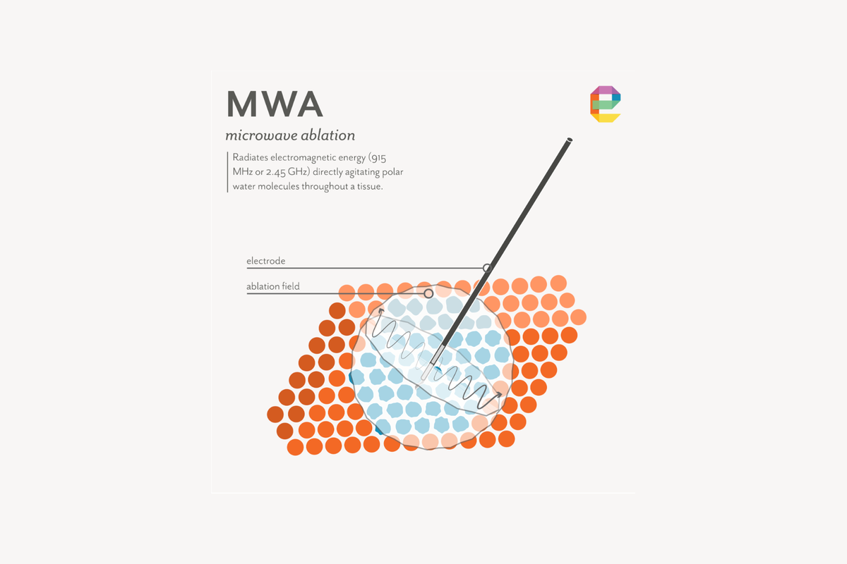
Microwave ablation (MWA) is a thermal ablation technology with a growing clinical footprint across liver, lung, kidney, bone, and a range of solid tumors. MWA works by delivering electromagnetic energy directly into tissue through an antenna embedded in a minimally invasive, needle-sized applicator. The target tissue is then heated to temperatures high enough to cause nearly instantaneous cell death.
The underlying physics will be familiar to anyone who has used a kitchen microwave oven to warm their food. Although many clinical MWA systems use the same operating frequency as a kitchen oven, the critical difference is the approach. A kitchen oven heats food from the outside in, radiating the food with microwave energy from all directions. An MWA applicator heats tissue from the inside out, radiating energy outward from an antenna within the target tumor.
That inside-out geometry is what lets MWA concentrate lethal temperatures within a defined volume of tissue while leaving surrounding structures safe. Understanding what MWA does to tissue, and what governs the size, shape, and predictability of the ablation zone, is the foundation for designing systems that perform reliably across the range of conditions they will encounter clinically.
This article summarizes the tissue effects of microwave ablation, drawn from our experience at Engenious Design developing microwave ablation systems, including novel directional applicators, bronchoscopic applicators, and generator platforms across multiple clinical applications.
Biological tissue contains many molecules that respond to an oscillating electric field: water is the most abundant and the strongest absorber at microwave frequencies, but proteins, lipids, and other polar molecules also contribute to the heating process. Water molecules are electrically asymmetric — the oxygen atom is slightly negative, and the hydrogen atoms are slightly positive — and therefore they act like tiny compass needles that want to flip orientation every time the externally applied electric field reverses direction.
At microwave frequencies, the field reverses billions of times per second. The molecules cannot keep up, and that lag between the field and the molecule generates friction. That friction generates heat.
This heating process, called dielectric heating, occurs throughout the volume of tissue exposed to the field, not just at a surface or electrode tip. That volumetric character distinguishes microwave ablation from radiofrequency ablation.
The amount of heat generated at any point in tissue depends on two factors: how strong the electric field is at that location, and how well that tissue absorbs microwave energy. These factors vary with tissue type, temperature and frequency. As a result, the MWA applicator design cannot be separated from the tissue it is meant to treat: an antenna tuned for liver may perform very differently in lung or bone.[1]
In radiofrequency ablation (RFA), the targeted tissue is part of the electrical circuit: current flows from the electrode, through tissue, and returns to a dispersive ground pad (or another electrode). As the tissue nearest the electrode heats and desiccates, its electrical resistance rises sharply, the circuit delivers less energy to the tissue, and growth of the ablation zone stalls. This impedance roll-off is the fundamental reason ablation zones in RFA are limited in size, and why RFA systems often use pulsed energy delivery to allow partial rehydration of tissue during treatment.
Microwave ablation does not work this way. The microwave field radiates outward from the antenna into tissue the way a radio signal radiates into air. Tissue resistance does not gate the delivery, because there is no resistive circuit the way there is in RFA.
How far and how effectively the field penetrates is determined by the tissue's ability to absorb and transmit microwave energy at the operating frequency, not by its local electrical resistance. This is why MWA can deliver power continuously at levels far above what RFA can sustain, why it can heat tissue with high electrical resistance like lung and bone that defeat RFA, and why it does not require pulsed delivery to manage the impedance ceiling that limits RFA.[2,3]
Water is the dominant absorber of microwave energy in tissue, so water content is the strongest predictor of how well a given tissue type heats. Soft organs (liver, kidney, muscle) and most solid tumors have high water content and absorb microwave energy readily. Fat has much lower water content and heats poorly. Proteins and other biomolecules contribute to absorption as well, which is why tissue composition as a whole — not just water fraction — determines the heating response.
Lung parenchyma, which is mostly air-filled, is a poor microwave absorber overall, but solid pulmonary tumors retain high water and protein content and heat more effectively relative to the surrounding aerated lung. This is an important practical advantage for microwave ablation in the lung.[3,4] Metastatic bone tumors may also heat much more effectively relative to surrounding healthy bone, which is a poor absorber due to its dense mineral structure and low water content.
As tissue heats, its ability to absorb microwave energy changes, and those changes feed back into the pattern of heating. This is one of the most important and least intuitive aspects of microwave ablation. At temperatures approaching 80 to 100°C, tissue water begins to evaporate, and proteins are fully denatured. As water is driven out and tissue structure breaks down, the tissue absorbs microwave energy less effectively in the region immediately adjacent to the applicator.[1] The practical effect is that the heating pattern shifts during the procedure: early on, heating concentrates near the antenna tip; as that tissue desiccates and loses its water, the microwave field penetrates further into wetter, intact tissue beyond. This is why ablation zone growth is not linear over time, and why device models that assume fixed tissue properties throughout the procedure are less accurate than those that account for these changes.[1,5]
The ablation zone in microwave ablation is the volume of tissue that reaches temperatures sufficient for irreversible cell death. That threshold is not a fixed number — it is a tradeoff between temperature and time. Tissue held at around 46°C for roughly an hour sustains irreversible damage, but that same outcome occurs in 4 to 6 minutes at 50–52°C, and in seconds at temperatures above 60°C.[2,6] In practice, clinical microwave ablation protocols aim to drive the entire target volume above 60°C to ensure rapid, reliable cell kill throughout, rather than relying on the slower kinetics that operate at lower temperatures. Temperatures above about 100°C cause tissue water to boil and vaporize, which can disrupt energy delivery and create unpredictable heat transfer effects in tissue.[1,2]
A fully developed microwave ablation treatment zone has a characteristic three-zone structure visible when the tissue is sectioned after treatment:
Standard linear antenna designs, like those used in microwave ablation applicators, radiate maximally in the radial direction (perpendicular to the antenna axis) and minimally along the axis itself. The resulting power absorption pattern is roughly toroidal (donut-shaped) around the antenna. However, most clinical microwave ablation applicators produce ellipsoidal ablation zones that are elongated along the applicator's axis, with the long axis running from the tip toward the handle. This is in part because the active length of the antenna can be as long as, or longer than, the absorption depth of microwaves in tissue, and because thermal conduction from heated tissue can extend the ablation zone axially.[5,8]
A further complication is the ablation "tail": heating along the outer conductor of the coaxial transmission line that feeds the antenna produces a zone of tissue heating proximal to the intended target, extending up the applicator shaft. This tail is a known artifact of unbalanced antenna designs and is mitigated in practice by active water cooling of the applicator shaft and by antenna design features (chokes, sleeves) that suppress current flow along the outer conductor. Despite these countermeasures, a residual tail is present in most commercial applicators and should be accounted for in treatment planning.[5,8]
One limitation of conventional MWA applicators is that their nearly cylindrically symmetric radiation pattern (as described above) often cannot be reliably positioned to avoid harming critical anatomy. If a tumor abuts the gallbladder, diaphragm, or the spinal cord, for example, the treating physician must either accept the risk of thermal injury to that structure, apply thermoprotective techniques (hydrodissection, gas dissection, endoluminal cooling) to create separation, or decline to treat.[9]
Directional microwave ablation (dMWA) applicators address this by integrating metallic reflector elements into the applicator shaft that restrict microwave radiation to a preferred angular half-space, producing a hemispherical rather than cylindrically symmetric ablation zone. Our team has experience developing experimental dMWA applicators that let the operator direct energy toward the tumor and away from structures that should not sustain thermal injury, an approach applied to adrenal and spinal indications in animal studies.[10,11]
MWA heats tissue based on its composition and the local field strength, without discriminating between cell types. A tumor cell and the wall of the bile duct next to it respond to the same field and reach the same temperature if they share the same location. Everything in the ablation zone that reaches lethal temperature is destroyed. This is both MWA's strength (reliable, complete cell kill within the treatment zone) and the core engineering challenge (keeping lethal temperatures confined to the target and away from structures that cannot be sacrificed).
The functional cells of the target organ (liver cells, kidney cells, lung cells, adrenal cells) are the intended targets of MWA and are reliably destroyed within the ablation zone. Cell death is complete within the central and main ablation zones. At the outer margin, cell kill becomes probabilistic, which is why treatment protocols target temperatures well above the minimum lethal threshold at the intended treatment boundary.[2,6]
Small vessels (below approximately 3 mm in diameter) within the ablation zone are typically sealed and destroyed by the heat. Larger vessels are more resistant to thermal injury for two reasons: their walls are thicker and more heat-tolerant, and blood flowing through the vessel continuously carries thermal energy away from the wall before it can accumulate to lethal levels.
This cooling effect of blood flow is a recognized limitation of MWA and can result in viable tumor tissue surviving immediately adjacent to a large vessel even when the surrounding tissue is completely destroyed.[3,7] This pattern of recurrence at the vessel margin is well documented in liver tumors treated near the major hepatic veins and is a common driver of incomplete local treatment in highly vascular target sites.
The thin inner lining of a bile duct or ureter can be damaged at temperatures lower than those required to destroy the surrounding tissue, and the duct wall, once thermally injured, may heal with permanent narrowing rather than full recovery. Narrowing of major bile ducts following MWA of centrally located liver tumors is a recognized complication that limits where thermal ablation can safely be applied.[9] Injecting saline or CO₂ between the duct and the ablation zone (a technique called hydrodissection or gas dissection) to create physical separation is standard practice in difficult cases, but adds steps and is not always feasible. Directional MWA can reduce the energy directed toward a duct, but cannot fully protect it when the tumor and duct are inseparable.[9,10]
Nerves are susceptible to both direct thermal destruction within the ablation zone and heat injury in the transition zone that can result in temporary or permanent loss of function. The temperature threshold for nerve damage is lower than for tissue destruction: temperatures above roughly 45°C held for several minutes can injure nerve fibers, and temperatures above 60°C cause irreversible structural damage.[6,13] This means the outer margin of an MWA treatment zone, which extends beyond the grossly visible zone, represents a risk zone for any nerve running nearby.[6,9] Applicator placement and treatment planning are the primary mitigations. Where critical nerves are very close to the treatment target (for example, the nerve that controls breathing during chest procedures, or nerves running between the ribs), hydrodissection or gas dissection is used to create physical separation.
MWA may be preferred over RFA for osseous metastases because high cortical bone impedance defeats RFA's current-based heating mechanism, while microwaves propagate through bone regardless of its resistance. However, treating vertebral metastases requires heating the lesion while protecting the spinal cord, which may be separated from the ablation target by only a few millimeters of bone.[11] Conventional symmetric MWA applicators cannot reliably hold that margin. Directional MWA, by confining the ablation zone to the anterior or lateral vertebral body, can spare the posterior elements and spinal canal.[11]
The tissue-level physics of MWA translate directly into the hardware and software decisions that determine whether a system performs reliably. A few of the most consequential engineering implications, from our experience designing MWA systems:
Microwave ablation destroys its clinical targets by radiating electromagnetic energy that converts to heat when absorbed in tissue. Ablation zone size and shape are determined by the antenna's radiation pattern, by tissue composition and how that composition changes as it heats, and by the applicator's internal cooling system.
Shaping the treatment zone to cover the entire target tumor, while keeping it away from structures that would harm the patient if they were thermally injured, is an engineering problem that starts at the antenna design stage and runs through generator architecture, treatment planning, and clinical protocol specification.
MWA has meaningful and well-documented limitations: the heat-sink effect from large vessels, the risk of collateral thermal injury to bile ducts and nerves, and the inability of conventional symmetric applicators to direct energy away from adjacent critical structures.
These are not reasons to avoid MWA. They are design requirements that define what a better MWA system looks like. Our team's work on directional MWA applicators, bronchoscopic delivery systems, and intraprocedural monitoring methods is aimed directly at those limitations.
Our team at Engenious Design has developed over 20 ablation systems across a range of energy modalities including MWA, RFA, and pulsed field ablation (PFA). If you are developing an MWA system and want an experienced engineering team on your side, contact us at connect@engenio.us.
1. Prakash P. Principles and technologies for electromagnetic-based tissue ablation. Engineering in Medicine and Biology Society, Academic Press (Elsevier). 2022.
2. Brace CL. Radiofrequency and microwave ablation of the liver, lung, kidney, and bone: what are the differences?. Curr Probl Diagn Radiol. 2009;38(3):135–143. doi:10.1067/j.cpradiol.2007.10.001
3. Lubner MG, Brace CL, Hinshaw JL, Lee FT. Microwave tumor ablation: mechanism of action, clinical results, and devices. J Vasc Interv Radiol. 2010;21(8 Suppl):S192–S203. doi:10.1016/j.jvir.2010.04.007
4. Sebek J, Kramer S, Rocha R, et al.. Bronchoscopically delivered microwave ablation in an in vivo porcine lung model. ERJ Open Res. 2020;6(4):00146–2020. doi:10.1183/23120541.00146-2020
5. Pfannenstiel A, Iannuccilli J, Cornelis FH, Dupuy DE, Beard WL, Prakash P. Shaping the future of microwave tumor ablation: a new direction in precision and control of device performance. Int J Hyperthermia. 2022;39(1):664–674. doi:10.1080/02656736.2021.1991012
6. Chu KF, Dupuy DE. Thermal ablation of tumours: biological mechanisms and advances in therapy. Nat Rev Cancer. 2014;14(3):199–208. doi:10.1038/nrc3672
7. Donlon P, Dennedy MC. Thermal ablation in adrenal disorders: a discussion of the technology, the clinical evidence and the future. Curr Opin Endocrinol Diabetes Obes. 2021;28(3):291–302. doi:10.1097/MED.0000000000000627
8. Fallahi H, Prakash P. Antenna designs for microwave tissue ablation. Crit Rev Biomed Eng. 2018;46(6):495–521. doi:10.1615/CritRevBiomedEng.2018028554
9. Garnon J, Cazzato RL, Caudrelier J, et al.. Adjunctive thermoprotection during percutaneous thermal ablation procedures: review of current techniques. Cardiovasc Intervent Radiol. 2019;42(3):344–357. doi:10.1007/s00270-018-2089-7
10. Pfannenstiel A, Sebek J, Fallahi H, et al.. Directional microwave ablation: experimental evaluation of a 2.45-GHz applicator in ex vivo and in vivo liver. J Vasc Interv Radiol. 2020;31(8):1270–1277. doi:10.1016/j.jvir.2020.01.016
11. Pfannenstiel A, Avellar H, Hallman C, et al.. Directional microwave ablation in spine: experimental assessment of computational modeling. Int J Hyperthermia. 2024;41(1):2313492. doi:10.1080/02656736.2024.2313492
12. Zeinali N, Pfannenstiel A, Hallman C, et al.. Transmission-based monitoring of dual-applicator microwave ablation discriminates discontiguous from contiguous ablation zones in an in vivo porcine liver model. J Vasc Interv Radiol. 2025;36(5):884–893.e3. doi:10.1016/j.jvir.2024.12.005
13. Haveman J, Sminia P, Wondergem J, van der Zee J, Hulshof MC. Effects of hyperthermia on the central and peripheral nervous systems: a review. Int J Hyperthermia. 2004;20(3):311–325. doi:10.1080/02656730310001637631