Minimally invasive electrical energy-based tissue ablation has transformed the management of a wide spectrum of clinical conditions, from solid cancer tumors to cardiac arrhythmias, from benign endocrine adenomas to symptomatic uterine fibroids. By delivering precisely controlled energy to a targeted tissue volume through a percutaneous or catheter-based approach, ablation can achieve outcomes comparable to open surgery with substantially reduced procedural burden, shorter recovery, and lower complication rates.
Three electrical energy modalities now dominate the clinical and device-development landscape: radiofrequency ablation (RFA), microwave ablation (MWA), and the newer non-thermal pulsed field ablation (PFA), also known as irreversible electroporation (IRE). While all three can destroy unwanted tissue, their underlying mechanisms lead to distinct clinical profiles with important advantages and limitations across both oncologic and non-oncologic applications. Understanding these differences is essential for device design, clinical decision-making, and regulatory strategy.
This practical article summarizes the differences, informed by our experience at Engenious Design where our team members have developed over 20 different ablation systems over the past 25 years.
Radiofrequency ablation (RFA)
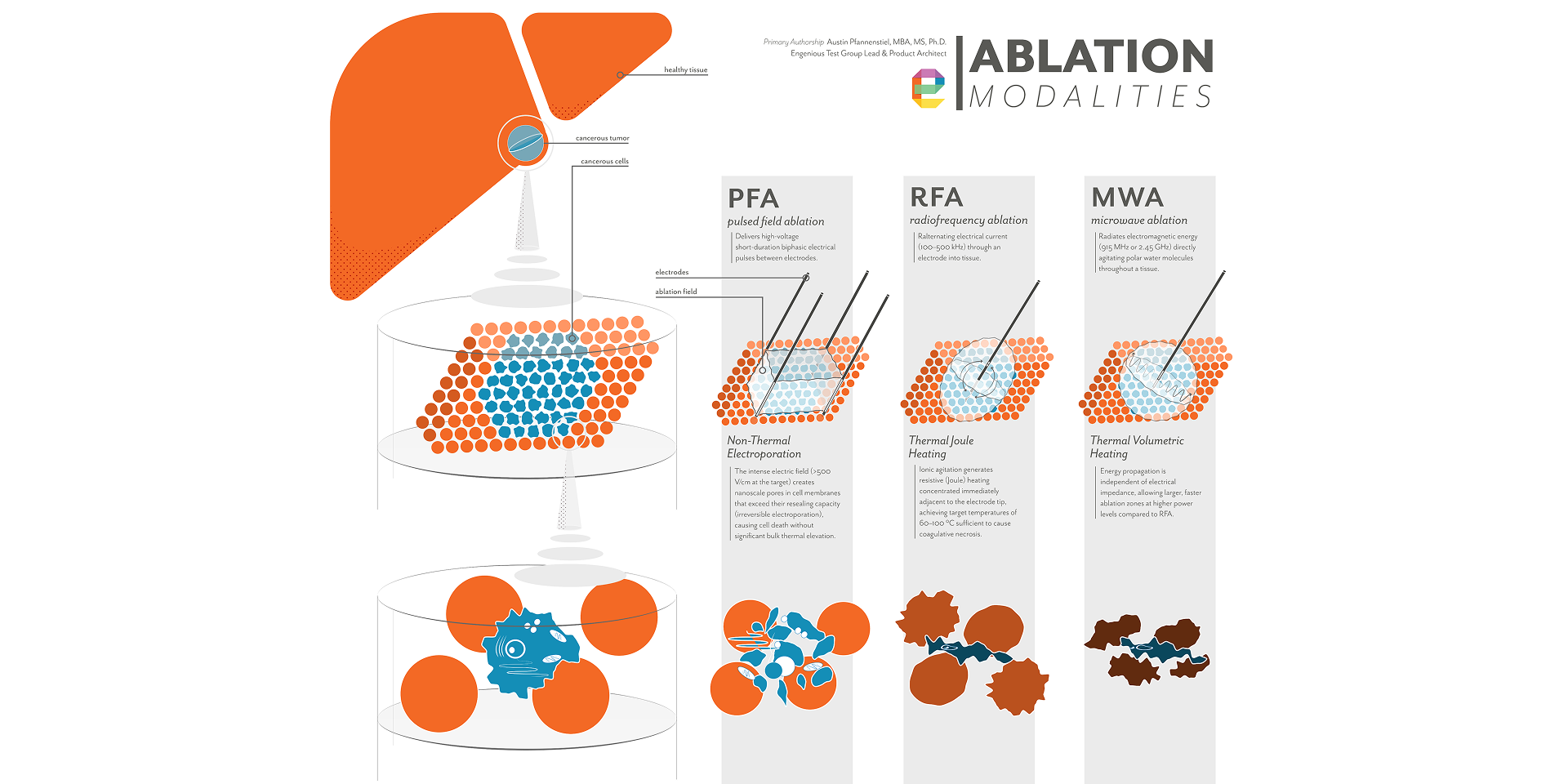
RFA delivers alternating electrical current (100–500 kHz) through an electrode into tissue. Ionic agitation generates resistive (Joule) heating concentrated immediately adjacent to the electrode tip, achieving target temperatures of 60–100 °C sufficient to cause coagulative necrosis. A fundamental limitation is impedance roll-off: tissue desiccation elevates electrical impedance and impedes further energy penetration, requiring pulsed delivery and restricting achievable ablation zone dimensions.[1] The heat-sink effect, convective cooling from adjacent large vessels, further reduces efficacy near vascular structures.[1,2] Electrode geometry, placement and other characteristics have a major impact on the resulting tissue effects. The majority of the Engenious team’s experience is with development of RFA applications.
Microwave ablation (MWA)
MWA radiates electromagnetic energy at 915 MHz or 2.45 GHz, directly agitating polar water molecules throughout a tissue volume. Energy propagation is independent of electrical impedance, allowing larger, faster ablation zones at higher power levels compared to RFA. MWA provides better tissue penetration, more predictable ablation geometry, shorter procedure times, and is less susceptible to the heat-sink effect than RFA.[1,3] However, cable heating necessitates active water or gas cooling, adding to applicator diameter and system complexity.[1]
The vast majority of commercially available MWA applicators produce near-spherical or ellipsoidal treatment zones symmetric around the applicator axis, limiting their ability to protect adjacent critical structures. Developmental directional MWA (dMWA) technology, using integrated metallic reflector elements to restrict microwave radiation to a preferred angular half-space, confers spatial control not available in conventional devices.[3,4] Engenious team members have unique experience with dMWA technologies.
Pulsed field ablation (PFA) / irreversible electroporation (IRE)
PFA delivers high-voltage, short-duration biphasic electrical pulses between needle electrodes. Rather than heating tissue, the intense electric field (>500 V/cm at the target) creates nanoscale pores in cell membranes that exceed their resealing capacity (irreversible electroporation), causing cell death without significant bulk thermal elevation. This non-thermal mechanism is the defining clinical differentiator: thermally sensitive structures including major blood vessels, bile ducts, ureters, and peripheral nerves are spared even when directly within the treatment field, because the structural collagen scaffold is preserved at doses lethal to parenchymal cells.[5,6] Engenious team members have significant experience developing PFA system, one of the field’s most active areas over the past eight years, and a trend we expect to continue.
PFA's non-thermal mechanism enables ablation of tissue adjacent to structures that are at prohibitive risk from heat-based modalities: hilar bile ducts in centrally located liver tumors, the ureteral collecting system in renal tumors, peripancreatic vasculature, the neurovascular bundle in prostate procedures, and perivascular conduction tissue in cardiac applications.[5,6]
Thermal modalities require thermoprotective techniques (hydrodissection, carbodissection, balloon interposition, or endoluminal cooling) to create physical separation between the ablation zone and vulnerable anatomy. These techniques add procedural complexity, are not universally feasible, and do not eliminate thermal risk when the target and critical structure share the same organ.[2]
Convective heat loss from vessels larger than approximately 3 mm reduces ablation efficacy. Examples include the hepatic veins and the portal triad, a well-documented cause of local tumor recurrence with RFA. Because PFA does not rely on heat accumulation, large vessels traversing the treatment field do not compromise ablation completeness. MWA partially mitigates the heat-sink effect through higher power delivery, but does not eliminate it entirely.[1,3]
We directly observe that RFA efficacy depends critically on tissue electrical impedance. Tissue desiccation from heating elevates impedance and limits energy penetration into surrounding tissue, necessitating pulsed delivery and restricting achievable ablation zone dimensions.[1] High-impedance environments such as cortical bone, previously desiccated tissue, or heterogeneous tumors particularly constrain RFA. Both MWA and PFA are impedance-independent, making them more consistently effective across diverse tissue types.
In our experience, conventional RFA and MWA create largely symmetric, ellipsoidal treatment zones. The vast majority of commercially available MWA applicators lack the consistency, predictability, and spatial control needed to treat targets near vulnerable structures.[3] Developmental directional MWA addresses this by restricting the ablation zone to a hemispherical region, demonstrated in liver and validated computationally and experimentally in spinal applications.[4,7] dMWA additionally enables a surface or extraluminal application mode, where the applicator does not penetrate the target organ, which is especially important for fragile glandular organs, such as the adrenal.[7,8]
PFA's zone of cell kill is determined by the electric field distribution between electrodes. Multi-electrode arrays allow customizable field shaping through selective activation and pulse sequencing.[5,6]
Both thermal modalities generate an immune response at the ablation margin. Immune infiltrates of monocytes/macrophages, neutrophils, dendritic cells, and lymphocytes have been documented in the transition zone following both RFA and MWA, and tumor-specific T-cell responses have been associated with improved recurrence-free survival.[1] PFA/IRE is believed to induce a qualitatively more robust immunogenic cell death: intact cellular components released during electroporation activate dendritic cells and drive tumor-specific T-cell responses in a manner potentially superior to the coagulative necrosis of thermal modalities. This immune activation is being explored in combination with checkpoint inhibitors in pancreatic and prostate cancer.[5,6]
.jpg)
From our experience on the front lines of development, we see that medical device engineers and development teams face the choice of energy modality which fundamentally shapes every layer of product architecture:
The three dominant electrical energy-based ablation modalities serve overlapping but distinct clinical niches across both oncologic and non-oncologic medicine. RFA, MWA and PFA/IRE.
Radiofrequency ablation remains the most established technology with the deepest evidence base, from hepatic tumors to cardiac arrhythmias to thyroid nodules, but is constrained by impedance roll-off, heat-sink sensitivity, and limited spatial control.[1,3]
Microwave ablation addresses several of RFA's thermal limitations through impedance-independent, high-power volumetric heating, and emerging directional MWA platforms represent a meaningful advance in spatial selectivity for applications where critical structures limit conventional thermal delivery.[3,4,7,8]
Pulsed frequency ablation/Irreversible electroporation offers a mechanistically distinct capability (non-thermal, selective cell death that preserves critical adjacent structures) that enables procedures not safely achievable with either thermal modality. PFA has already transformed the cardiac electrophysiology landscape and is positioned to do the same across a growing range of solid-organ oncologic and benign applications.[5,6,10,11]
For device developers like ourselves, each modality presents distinct engineering challenges and differentiated commercial opportunities. The continued maturation of PFA across organ systems, the emerging precision of directional MWA, and the expanding role of ablation in benign disease management collectively define an innovation landscape with significant unmet clinical needs and substantial room for purposeful engineering to advance patient outcomes.
Developing an ablation system? Contact Engenious Design to see how our team’s 25 years of experience across ablation technologies can help.
1. Donlon P, Dennedy MC. Thermal ablation in adrenal disorders: a discussion of the technology, the clinical evidence and the future. Curr Opin Endocrinol Diabetes Obes. 2021;28(3):291–302. doi:10.1097/MED.0000000000000627
2. Garnon J, Cazzato RL, Caudrelier J, et al. Adjunctive thermoprotection during percutaneous thermal ablation procedures: review of current techniques. Cardiovasc Intervent Radiol. 2019;42(3):344–357. doi:10.1007/s00270-018-2089-7
3. Pfannenstiel A, Iannuccilli J, Cornelis FH, Dupuy DE, Beard WL, Prakash P. Shaping the future of microwave tumor ablation: a new direction in precision and control of device performance. Int J Hyperthermia. 2022;39(1):664–674. doi:10.1080/02656736.2021.1991012
4. Pfannenstiel A, Sebek J, Fallahi H, et al. Directional microwave ablation: experimental evaluation of a 2.45-GHz applicator in ex vivo and in vivo liver. J Vasc Interv Radiol. 2020;31(8):1270–1277. doi:10.1016/j.jvir.2020.01.016
5. Davalos RV, Mir LM, Rubinsky B. Tissue ablation with irreversible electroporation. Ann Biomed Eng. 2005;33(2):223–231. doi:10.1007/s10439-005-8981-8
6. Scheffer HJ, Nielsen K, de Jong MC, et al. Irreversible electroporation for nonthermal tumor ablation in the clinical setting: a systematic review of safety, efficacy, and outcomes. J Vasc Interv Radiol. 2014;25(7):997–1011. doi:10.1016/j.jvir.2014.01.028
7. Pfannenstiel A, Avellar H, Hallman C, et al. Directional microwave ablation in spine: experimental assessment of computational modeling. Int J Hyperthermia. 2024;41(1):2313492. doi:10.1080/02656736.2024.2313492
8. Donlon PT, Fallahi H, Beard WL, et al. Using microwave thermal ablation to develop a subtotal, cortical-sparing approach to the management of primary aldosteronism. Int J Hyperthermia. 2019;36(1):905–914. doi:10.1080/02656736.2019.1650205
9. Sebek J, Kramer S, Rocha R, et al. Bronchoscopically delivered microwave ablation in an in vivo porcine lung model. ERJ Open Res. 2020;6(4):00146–2020. doi:10.1183/23120541.00146-2020
10. Reddy VY, Anter E, Rackauskas G, et al. Pulsed field ablation for pulmonary vein isolation in paroxysmal atrial fibrillation: 1-year outcomes from the IMPULSE, PEFCAT, and PEFCAT II studies. JACC Clin Electrophysiol. 2021;7(5):614–627. doi:10.1016/j.jacep.2021.02.014
11. Duytschaever M, De Potter T, Weerasooriya R, et al. Pulmonary vein isolation with a pulsed-field ablation system: 1-year outcomes from the MANIFEST-PF registry. Europace. 2023;25(1):316–327. doi:10.1093/europace/euac153
12. Liu SY, Chu CM, Kong AP, et al. Radiofrequency ablation compared with laparoscopic adrenalectomy for aldosterone-producing adenoma. Br J Surg. 2016;103(10):1476–1486. doi:10.1002/bjs.10226
13. Lim HK, Lee JH, Ha EJ, et al. Radiofrequency ablation of benign non-functioning thyroid nodules: 4-year follow-up results for 111 patients. Eur Radiol. 2013;23(4):1044–1049. doi:10.1007/s00330-012-2671-3
14. Diao Z, Liu X, Qian L, et al. Efficacy and its predictor in microwave ablation for severe secondary hyperparathyroidism in patients undergoing haemodialysis. Int J Hyperthermia. 2016;32(6):614–622. doi:10.1080/02656736.2016.1191079
This article was prepared for informational and educational purposes. Clinical decisions should be based on current peer-reviewed evidence and applicable regulatory guidance.